|
||||||||||||
 |
 |
 |
 |
 |
 |
 |
 |
| 购买进口仪器、试剂和耗材——就在始于2001年的毕特博生物 www.bitebo.com |
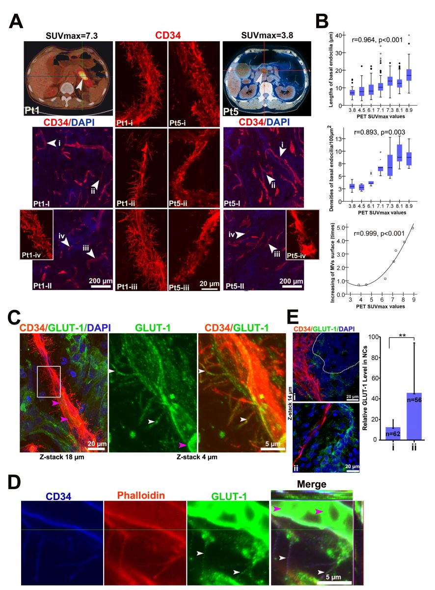
胰腺癌是一个几乎没有治疗的恶性肿瘤,得了胰腺癌等于判死刑,一半病人会六个月内死去,故称之为“癌症之王”。 胰腺位于腹腔深部位置和胰腺癌症状不明显,常规的影像学检查很难发现早期胰腺癌,因此早期发现或诊断胰腺癌很难。这种肿瘤进展凶猛、致死性极强、对大部分化疗药物治疗敏感,而且手术切除率低。当胰腺癌被发现时,大部分病人肿瘤已经到中晚期。 胰腺癌有非常厚的“堡垒”,即大量的细胞外间质和极度贫乏的血管,但胰腺癌具有超强的营养物质获取能力。赛音贺西格老师与中山医院楼文辉教授合作,通过七年的不懈努力,建立了高分辨率的3D重建肿瘤内部结构染色方法。通过这种方法揭开了胰腺癌的神秘物质运输通道的面纱-发现一种新型的“毛毛”微血管。这些微血管的细胞的背面长出巨大的突起,使微血管血管变成一个大量突起的类似巨型“毛毛虫”的怪物。 临床标本的数据分析结果显示血管内皮细胞基底面微绒毛可能大大提高了微血管的糖运输能力。他们把这个新发现的内皮细胞突起命名为“Basal microvilli "-内皮细胞基底微绒毛 ",并把这个发现发表在国际顶尖病理学杂志“The Journal of pathology ",美国北卡大学的Andrew C. Dudley and Victoria L. Bautch博士同期发表了“Feeding cancer’s sweet tooth: specialized tumor vasculature shuttles glucose in Pancreatic ductal adenocarcinoma”-“喂肿瘤的糖牙-胰腺导管癌内糖运输的血管快车”评论文章。这些细胞突起深入到胰腺癌组织的发达的间质中,甚至可以延伸到肿瘤细胞周围或肿瘤细胞之间。如同小肠微绒毛,血管内皮细胞基底面微绒毛大大增加了微血管内皮细胞的交换面积和物质交换深度,从而极少微血管可以满足胰腺癌细胞旺盛的代谢需求。 虽然体内有些细胞,例如骨细胞,星型胶质细胞等,通过细胞突起获得能量物质,但在极性的上皮细胞当中一般不存在基底面具有物质交换功能突起,这是首次观察到血管内皮基底面存在大量具有物质交换功能的突起。这种特殊的细胞突起只有在肿瘤内存在,这点可能是胰腺癌的致命弱点-“Achilles heels”。 利用这个胰腺癌特异的途径输送治疗药物或者通过靶向分子抑制或破坏此结构,可能将成为治疗胰腺癌的或首选途径。此项工作得到余龙教授和很多国内外同行大力支持。 Pancreatic ductal adenocarcinoma (PDAC) is a nearly lethal neoplasm. It is a remarkably stroma-rich, vascular-poor and hypo-perfused tumour, which prevents efficient drug delivery. Paradoxically, the neoplastic cells have robust glucose uptake, suggesting that the microvasculature has adopted an alternative method for nutrient uptake and cellular trafficking. Using adapted thick tumour section immunostaining and three-dimensional (3D) construction imaging in human tissue samples, we identified an undiscovered feature of the mature microvasculature in advanced PDAC tumours; long, hair-like projections on the basal surface of microvessels that we refer to as 'basal microvilli'. Functionally, these basal microvilli have an actin-rich cytoskeleton and endocytic and exocytic properties, and contain glucose transporter-1 (GLUT-1)-positive vesicles. Clinically, as demonstrated by PET–CT, the tumour microvasculature with the longest and most abundant basal microvilli correlated with high glucose uptake of the PDAC tumour itself. In addition, these basal microvilli were found in regions of the tumour with low GLUT-1 expression, suggesting that their presence could be dependent upon the glucose concentration in the tumour milieu. Similar microvasculature features were also observed in a K-Ras-driven model of murine PDAC. Altogether, these basal microvilli mark a novel pathological feature of PDAC microvasculature. Because basal microvilli are pathological features with endo- and exocytic properties, they may provide a non-conventional method for cellular trafficking in PDAC tumours. |
购买进口仪器、试剂和耗材——就在始于2001年的毕特博生物
www.bitebo.com |
|